
The Brain Was Always Two — Stanford Just Shattered Decades of Textbook Assumptions
Summary
A September 2026 study published in Nature Neuroscience by a Stanford Medicine-led team reveals that the front and back of the developing brain arise from two entirely distinct progenitor cell lineages — Otx2-expressing cells independently building the forebrain and midbrain while Gbx2-expressing cells independently build the hindbrain — with this separation established from gastrulation onward and locked in by fundamental chromatin architecture differences. This finding overturns the long-standing developmental biology assumption that a single progenitor population generates the entire brain, and it explains why decades of attempts to derive hindbrain neurons by redirecting forebrain progenitors consistently produced nothing useful. Building on this corrected developmental logic, the team became the first to successfully derive functional hindbrain motor neurons from human pluripotent stem cells — neurons that generate action potentials and express segmental markers associated with the control of facial, swallowing, and throat musculature. The practical significance of this new tool extends to spinal muscular atrophy and ALS, both of which involve hindbrain motor neuron loss that drives life-threatening swallowing and respiratory complications, as well as to understanding how weight-loss drugs like semaglutide act via hindbrain appetite circuits. Evolutionary analysis confirmed the two-origin pattern across chicken, zebrafish, and acorn worms — species whose lineages diverged from ours roughly 550 million years ago — suggesting that this dual-progenitor architecture is a deeply conserved feature of animal nervous system development. Beyond its empirical contributions, the study offers a methodological object lesson that travels far beyond neuroscience: the word "brain," by implying a single continuous organ with a singular origin, constrained a generation of experimental design, and recognizing that constraint turned out to be as important as any molecule the team characterized.
Key Points
Two Separate Progenitor Lineages Emerging From the Very Start of Development
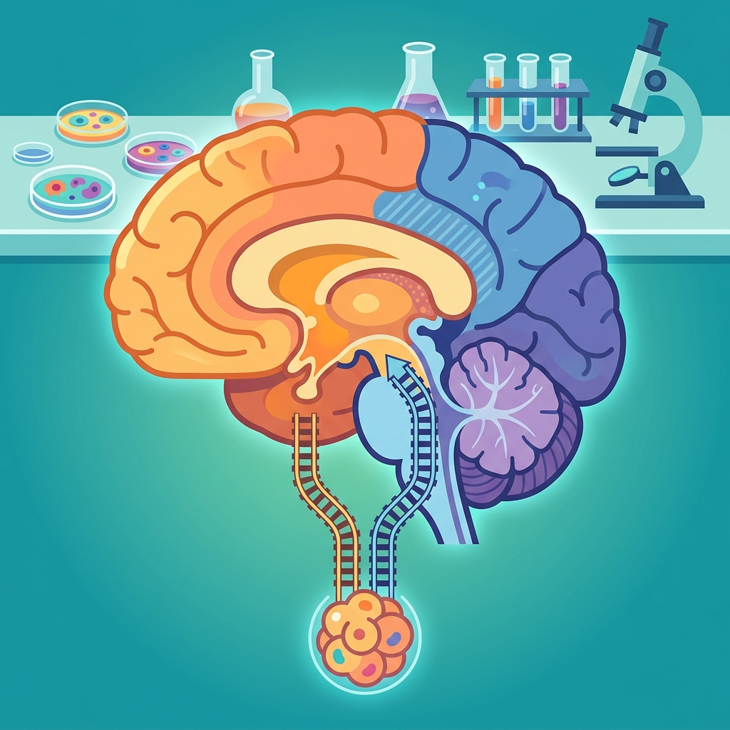
The conventional story of brain development held that a single neural progenitor population gives rise to all three major brain regions — forebrain, midbrain, and hindbrain — through progressive differentiation from a shared origin. The Stanford team's mouse embryo lineage-tracing experiments directly challenged this model. Otx2-expressing cells, which build the forebrain and midbrain, and Gbx2-expressing cells, which build the hindbrain, emerge simultaneously during gastrulation — the earliest stage of embryonic organization — as two completely separate, never-overlapping populations. Senior author Kyle Loh summarized it plainly: "We've shown for the first time that the front of the brain arises from a totally different progenitor cell than the back of the brain." The significance here is not merely that two cell types exist side by side — it's that they were never part of the same progenitor pool to begin with. The posterior neural ectoderm did not branch off from the anterior; they ran on parallel tracks from the start. This forces a fundamental revision to how the developmental origin of brain regions is understood, and it means any experimental protocol that began by trying to convert forebrain progenitors into hindbrain cells was built on a false premise from its first day.
Chromatin Architecture Differences Lock Each Lineage Onto Its Own Developmental Track
One of the most technically consequential aspects of the study is the discovery that chromatin architecture — the physical packaging of DNA — differs fundamentally between anterior and posterior neural ectoderm. Chromatin structure is not merely a readout of which genes are currently active; it encodes which genomic regions are accessible for future activation and which are structurally closed off, effectively pre-shaping the futures a cell can access. The fact that these differences are already established at the gastrulation stage means the developmental destinies of these cells are baked into their epigenetic architecture, not simply guided by local signaling environments that could be overridden. Loh described this as travelers on parallel tracks who cannot switch lines: once on their track, cells are committed. The mutual exclusivity of Otx2 and Gbx2 expression had been documented since the late 1990s — in work by Wassarman in 1997 and by Millet and Broccoli in 1999 — but those classical studies characterized a regional boundary, not a fundamental lineage split with distinct chromatin states dating to gastrulation. What Stanford added is mechanistic depth: not just "these cells express different transcription factors" but "these cells inhabit fundamentally different epigenetic states that prevent lineage crossing, period." That distinction is the foundation for everything that follows.
The First Successful Derivation of Hindbrain Motor Neurons From Human Stem Cells
The most immediately practical outcome of this research is the first successful derivation of functional hindbrain motor neurons from human pluripotent stem cells. These neurons generated action potentials and expressed segmental marker proteins associated with the control of facial muscles, swallowing, and throat function. Understanding this achievement requires an important clarification: making motor neurons from stem cells is not itself new. Wichterle and colleagues demonstrated spinal cord motor neuron derivation from embryonic stem cells in a 2002 Cell paper, and those spinal cord motor neurons have served as a standard ALS research tool for over two decades. The new achievement is specifically the derivation of hindbrain motor neurons — the population governing swallowing, speech, and breathing — which are anatomically and functionally distinct from spinal cord motor neurons and had never been successfully made from human stem cells before. Jokhai explained why previous attempts failed: "Previous attempts to make hindbrain neurons likely tried to coax forebrain and midbrain progenitors into hindbrain cells, which our study shows is not possible." The correct starting cell — posterior neural ectoderm — was finally identified, and using it enabled what had been impossible. Jokhai added: "Now we have a model to better understand these devastating diseases, and work toward regenerative therapies for them."
Evolutionary Conservation Spanning Roughly 550 Million Years of Animal History
The research team's finding didn't stop with mice and human stem cells. They confirmed the same two-origin pattern in chicken, zebrafish, and acorn worms — organisms representing roughly 550 million years of evolutionary divergence from the human lineage. Deep conservation of a developmental mechanism across that span of time is not a trivial observation. When the same architectural principle persists through hundreds of millions of years of animal evolution, it suggests the dual-lineage system is not a quirk of mammalian neurodevelopment but something closer to a foundational constraint on how animal nervous systems are assembled. Loh's interpretation is worth noting: "Our research suggests that evolution took two existing neural systems and pushed them together spatially" — meaning the unified brain we carry today may be the product of ancient, originally independent neural systems that were merged into proximity over evolutionary time. The observation that jellyfish, which diverged from our ancestors roughly 600 to 700 million years ago, maintain two separate nervous systems at opposite ends of their bodies is consistent with this reading. Loh used "suggests" rather than any stronger claim, which is precisely the right level of epistemic caution for a hypothesis of this scope.
How the Word "Brain" Functioned as a Multi-Decade Cognitive Constraint
The study's deepest contribution may be epistemological rather than purely empirical. Co-first author Jokhai identified the core problem with unusual clarity: "the word 'brain' implies a contiguous organ that likely has a singular origin." That implication is not a trivial semantic observation — it is a description of how scientific framing shapes scientific practice. In research, naming an object shapes which hypotheses feel natural, which experimental designs seem obvious, and how results get interpreted. "The brain" as a unitary concept made it natural to hypothesize a unitary progenitor pool, which made it natural to try redirecting progenitors across the front-back boundary, which is exactly what this paper shows was never going to work. The roughly thirty years between the characterization of the Otx2/Gbx2 boundary in the 1990s and this paper's conclusion that the boundary reflects separate lineages is, at least in part, the gap created by a naming convention that ruled out the right question. Classification systems are tools that open certain possibilities and close others, and this case illustrates the closing side with unusual sharpness.
Positive & Negative Analysis
Positive Aspects
- A Research Tool That Simply Did Not Exist Before
The most tangible benefit this study delivers to science is a tool that had no previous existence. Brainstem tissue cannot be biopsied from living patients — the surgical risk is prohibitive and the research justification insufficient — which meant there was no direct way to study what human hindbrain motor neurons do or how they fail in disease. Previous efforts to make these neurons starting from forebrain progenitors consistently failed for reasons nobody fully understood until now. The Gbx2-positive posterior neural ectoderm is the correct starting point, and establishing that starting point makes the derivation protocol principled and reproducible rather than improvisational. This is not a marginal refinement of an existing capability; it is the creation of a capability that opens up research questions that were structurally closed before. I think the significance of having the right starting cell — rather than working around the wrong one — is hard to overstate for a field that spent decades not understanding why its methods weren't producing results.
- New Research Pathways for SMA and ALS
Both spinal muscular atrophy and ALS involve progressive motor neuron loss, and both ultimately kill through the same mechanism: failure of the hindbrain motor neurons that control swallowing and breathing leads to aspiration pneumonia and respiratory collapse. SMA is the leading genetic cause of infant mortality in the first year of life. ALS primarily affects adults diagnosed between their forties and seventies, with the CDC National ALS Registry estimating approximately 34,720 affected individuals in the United States as of August 2026. Based on 2016 figures from the Korean ALS Association, around 2,000 to 2,500 patients were living with the disease in South Korea, with an estimated 400 to 500 new cases per year. A 2022 review by Jiang and colleagues found that the approved drugs of that period extended survival by only a matter of months, and three investigational drugs failed at Phase 3 during just the 2020 to 2022 window surveyed. The availability of human hindbrain motor neurons as a disease model creates, for the first time, a direct way to observe how SMA and ALS damage the specific neurons whose loss causes the most harm to patients.
- A New Window Into How Weight-Loss Drugs Like Semaglutide Work
One of the less immediately obvious applications of this research is its potential contribution to understanding the hindbrain circuits that GLP-1 receptor agonists like semaglutide act upon. The Stanford Medicine press release explicitly connected the work to the question of "precisely how weight-loss drugs like semaglutide work." A 2024 Nature paper by Huang and colleagues mapped the relevant circuitry, finding that GLP-1 receptor neurons in the Area Postrema drive appetite suppression paired with nausea, while GLP-1 receptor neurons in the nucleus tractus solitarius produce satiety without the nausea effect. Pharmacologically separating those two pathways could produce drugs that preserve the appetite-suppressing benefit of current GLP-1 agonists while substantially reducing the nausea and vomiting that many patients experience. Testing that kind of hypothesis requires making these specific hindbrain neurons from human cells and running drug candidates directly against them — which is the capability this research provides for the first time. Given the global scale of obesity as a public health challenge, the potential reach of this research thread may ultimately involve far more people than the neurodegenerative disease applications.
- A Methodological Object Lesson That Travels Beyond Neuroscience
This study does something rare: it demonstrates, in a concrete and falsifiable way, how a classification system can constrain scientific progress for decades without anyone necessarily doing bad science. The lesson is not specific to developmental neurobiology. Any scientific discipline that names phenomena in ways carrying hidden assumptions about their unity, origin, or mechanism runs a version of the same risk — the name does cognitive work that no individual experiment is equipped to undo. The finding invites researchers in other domains to ask whether any of their own foundational categories are performing similar invisible work, quietly ruling out hypotheses before they can be formed. I think this kind of methodological reflexivity is genuinely valuable, and it tends not to show up in citation counts or drug approvals — but it shapes the direction of inquiry over a generation. The fact that Loh's lab came from outside classical developmental neuroscience and saw what insiders had normalized away is itself a data point about the value of cross-disciplinary perspective in generating this kind of insight.
Concerns
- The Core Experiments Were Conducted in Mouse Embryos
The primary experimental work behind this study was performed in mouse embryos, and translation to human biology, while strongly suggested, has not been directly confirmed. The team extended findings to chicken, zebrafish, and acorn worms, broadening the evidential base considerably, and the human pluripotent stem cell experiments offer meaningful indirect support for human applicability. However, whether the specific timing and mechanism of gastrulation-stage lineage commitment observed in mice appears in the same form and at the same developmental moment in human embryos remains unverified by direct experimentation. Mouse and human embryogenesis differ in both developmental timeline and the specifics of gene regulatory networks, and these differences could modify the mechanism even if the general principle holds. Given the ethical constraints on human embryo research, direct verification is a difficult and slow process. Until it is complete, interpreting this finding as confirmed human biology rather than a well-supported and highly plausible hypothesis requires a degree of caution that media coverage has not always maintained.
- The "Two Organs" Label Risks Replacing One Misconception With Another
There is a genuine irony in the way this finding has been communicated publicly. The paper's central philosophical point is that the word "brain" — by implying a single continuous structure with a single origin — constrained scientific thinking for decades. But the popular framing that has emerged in response, calling the brain "two separate organs," introduces its own distortion. Anatomically, the brain is a continuous structure; what is "two" is its developmental origin. The label "two organs" implies physical separation that isn't there, and it could mislead students, patients, and researchers who encounter the claim secondhand. ScienceDaily's choice to include "may actually be" in their headline shows editorial awareness of the issue, but hedges tend to be stripped out as findings travel further from their source. I'd much rather see the standard formulation become "a brain with two developmental origins" — a description that is accurate, interesting, and does not create a new false impression to replace the old one.
- No Independent External Replication or Critique Is Yet Available
As of the time of writing, no independent research group has published a replication or critique of this study's findings in the open scientific record. All media coverage draws on statements from the Stanford team, and the peer review that led to Nature Neuroscience publication represents rigorous expert scrutiny but is not the same as independent replication. Peer review assesses whether a study's methods and conclusions are internally sound and credibly supported; independent replication asks whether the finding appears when a different group applies different tools in a different laboratory. For a claim of this magnitude — that a foundational assumption of developmental neurobiology requires revision — independent replication from multiple groups is the standard of evidence that should be required before treating it as settled. This is not a criticism of the paper's quality; it is a statement of what the scientific process demands before a single study, however well-executed and however prestigious its venue, graduates from finding to consensus.
- The Distance From Research Tool to Clinical Benefit Is Very Long
The creation of hindbrain motor neurons in the lab is a meaningful scientific milestone, but it would be a serious mistake to read this as a near-term therapeutic advance for ALS, SMA, or any other disease. There is an enormous distance between the ability to make a cell type in a dish and the ability to use that cell type to identify a treatment that genuinely helps patients. A 2022 review by Jiang and colleagues documented how difficult ALS drug development remains even with existing tools: among the investigational drugs entering clinical trials in the 2020 to 2022 window alone, three failed at Phase 3 after showing earlier-stage promise. The history of neurodegenerative disease drug development is a long record of promising tools requiring decades of refinement before yielding clinical impact. The new hindbrain motor neuron model will make certain experiments newly possible, and those experiments may eventually point toward better treatments — but I think it is important to say clearly, especially for patients and families living with SMA or ALS, that this research opens a door, it does not walk through it.
Outlook
The most immediate thing to watch, in the months following this paper's September 2026 publication in Nature Neuroscience, is whether independent research groups can replicate the core findings. The developmental biology and stem cell communities have taken note, and the critical question is whether the Otx2/Gbx2 lineage separation during gastrulation holds up across different mouse strains and different lineage-tracing technologies. I expect this part of the finding to pass replication without major difficulty. The reason is that the Otx2/Gbx2 boundary itself has been established for decades; what the Stanford team added was the demonstration that this boundary reflects chromatin-level lineage separation dating to gastrulation rather than a simple regional patterning event. The more demanding test will be whether other labs can reproduce the hindbrain motor neuron derivation protocol from human pluripotent stem cells. Stem cell differentiation protocols are notoriously sensitive to subtle differences in culture conditions, batch variation in growth factors, and laboratory-specific handling procedures. Getting consistent results for a genuinely novel cell type across multiple independent settings will require systematic effort and likely some iteration on the protocol itself.
A second short-term development worth tracking is the science communication debate around the "two organs" framing. That phrase is already circulating in mainstream media, and it's likely to draw pushback from developmental biologists who will insist on a clear distinction between "two developmental origins" and "two anatomically separate organs." I think that pushback will be healthy and necessary. The deeper question it raises — how much simplification in science communication crosses into distortion — is not unique to this paper, and this case gives the field a concrete example to reason about. ScienceDaily's choice to hedge its headline with "may actually be" signals some editorial awareness of the issue, but hedges tend to drop out as findings travel further from the original source. Science communication has a long memory, and the way this result gets encoded in popular understanding will shape how follow-up research is framed, funded, and received.
On the practical side, the hindbrain motor neuron derivation protocol will begin moving into stem cell laboratories around the world. Given that the research received funding from the SMA Foundation and HHMI, among other organizations, there are strong institutional incentives for broad protocol sharing. The SMA research community in particular is likely to move quickly, because SMA is the leading genetic cause of infant death in the first year of life, and the disease already has approved disease-modifying treatments — nusinersen, onasemnogene abeparvovec, and risdiplam — which means a functional pipeline between basic research and clinical application already exists. Whether the protocol proves consistently reproducible across different laboratory settings, and whether the resulting cells behave reliably as a disease model, are distinct technical challenges that will need to be addressed before the tool can be widely adopted.
Moving into the medium-term window of roughly six months to two years, the most anticipated development is the research team's own stated next step: studying how SMA and ALS damage hindbrain neurons. This was never previously a question with a direct experimental answer, because the correct cell type simply did not exist as a research tool. Consider what becomes newly possible: ALS affects both forebrain and hindbrain neurons — upper motor neurons in the cortex and lower motor neurons in the brainstem — and if those two populations arose from fundamentally different developmental lineages, the molecular mechanisms driving their degeneration may not be identical. If forebrain-lineage and hindbrain-lineage motor neurons break down through different pathways, ALS may need to be reconceptualized as a condition in which two developmentally distinct neuronal populations fail in their own ways. This possibility strikes me as quite real, and it is consistent with the long-standing clinical observation that ALS patients vary substantially in where their symptoms begin and how the disease progresses.
The semaglutide and GLP-1 receptor agonist research space is another medium-term area where the payoff from this work could be significant. The Stanford press release explicitly connected the research to understanding how weight-loss drugs like semaglutide work via hindbrain circuits. The 2024 Nature paper by Huang and colleagues identified that GLP-1 receptor neurons in the Area Postrema and the nucleus tractus solitarius serve distinct functions in the hindbrain: one pathway drives appetite suppression paired with nausea, while the other produces satiety without nausea. Selectively targeting the satiety pathway could, in principle, produce next-generation obesity treatments that preserve therapeutic benefit while eliminating the most common side effects. Testing that hypothesis requires the ability to make these specific hindbrain neurons from human stem cells and run drug candidates against them directly — which is exactly what this research makes newly possible. Given that obesity affects hundreds of millions of people globally, the scale of potential benefit from this research thread may ultimately exceed the neurodegenerative disease applications.
A third medium-term question the Stanford team explicitly flagged is the developmental origin of the spinal cord. If the spinal cord also turns out to arise from a progenitor lineage distinct from both anterior and posterior neural ectoderm, the developmental map of the entire central nervous system would need to be substantially redrawn. I think the right posture is to hold this as a genuinely open question. The answer matters directly for spinal cord injury research, where the question of which starting cell produces the right spinal cord neurons mirrors exactly what just happened in hindbrain research. If the same foundational error — using the wrong progenitor — has been made in spinal cord stem cell work, the methodological lesson from this Stanford paper would apply again in a new domain.
Looking further out, to the two-to-five year window, I think the most consequential shift this research could produce is a change in how introductory developmental neuroscience is taught. The statement that "a single progenitor population gives rise to the entire brain" would need to be replaced by something like "two independent progenitor lineages — anterior and posterior neural ectoderm — emerge in parallel from gastrulation onward." That replacement is not merely a sentence edit; it alters the conceptual architecture of everything that follows — the signaling networks, the disease models, the experimental logic. That kind of textbook revision requires multiple rounds of independent replication and broad field consensus, which realistically puts it in the three-to-five year range at earliest. But if the finding holds up across independent labs, I think the version of a developmental neuroscience textbook published in the early 2030s will look meaningfully different from today's on this foundational question.
A longer-range possibility — clearly speculative at this point, but worth naming — is the emergence of developmental-origin-based subtyping in neurodegenerative disease classification. Currently, diseases like ALS are classified primarily by protein pathology or clinical presentation. If the developmental lineage of affected neurons turns out to influence disease trajectory or drug response, origin-based classification could become a new axis in precision neurology. For instance, if forebrain-lineage upper motor neuron degeneration and hindbrain-lineage lower motor neuron degeneration respond differently to the same drug in ALS, that would have direct implications for clinical trial design and eventually for treatment selection. This scenario requires far more follow-up research than currently exists, and I want to be clear that I'm presenting it as a possibility worth investigating, not a prediction. But I think it's the kind of question this paper now makes it legitimate to ask.
Mapping this out as a scenario analysis: in an optimistic scenario, independent replication proceeds quickly, the hindbrain neuron protocol standardizes across labs, and meaningful new therapeutic targets emerge within a few years for SMA and ALS. In a base-case scenario, the two-origin finding itself replicates cleanly but clinical translation follows the usual long arc of basic research — the hindbrain motor neuron model becomes a valuable tool that gradually shifts the research landscape without producing dramatic near-term breakthroughs. In a pessimistic scenario, the human embryo validation proves more complex than the mouse data suggested, or the "two organs" media framing generates excessive expectations that the research timeline can't meet, leading to a premature pullback of attention and funding before the tool has been fully exploited. My honest assessment is that the base case is most likely. But regardless of how the therapeutic applications develop, I think the real legacy of this paper may rest in its methodological lesson: when decades of careful work keep failing to produce a result, one of the most productive places to look is the assumption embedded in the very name of the thing you're studying.
Sources / References
- Human brain is two separate organs, Stanford Medicine-led research finds — Stanford Medicine News (2026-09-18) — Human brain is two separate organs, Stanford Medicine-led research finds
- Two parallel neural ectoderm progenitors contribute to the developing brain — Nature Neuroscience (2026-09-18) — Two parallel neural ectoderm progenitors contribute to the developing brain
- Stanford scientists discover the human brain may actually be two separate organs — ScienceDaily (2026-09-21) — Stanford scientists discover the human brain may actually be two separate organs
- New developments and opportunities in drugs being trialed for amyotrophic lateral sclerosis from 2020 to 2022 — Frontiers in Pharmacology / PMC (Jiang et al. 2022) — New developments and opportunities in drugs being trialed for amyotrophic lateral sclerosis from 2020 to 2022
- National ALS Registry Dashboard — CDC (updated August 2026) — National ALS Registry Dashboard
- Dissociable hindbrain GLP1R circuits for satiety and aversion — Nature / PMC (Huang et al. 2024) — Dissociable hindbrain GLP1R circuits for satiety and aversion
- The Brain Is Two Separate Organs Joined by Evolution — NeuroscienceNews.com — The Brain Is Two Separate Organs Joined by Evolution